Difference between revisions of "File:BiocellionWiki Wrinkleformation.png"
From Biocellion
| Line 1: | Line 1: | ||
| − | Sequence of wrinkle formation originating from cell death at the cell-agar substratum interface. | + | Sequence of wrinkle formation originating from cell death at the cell-agar substratum interface. Left column shows cross-sectional images of a wrinkle from [1], green color shows the area of cell death, while the right column shows the simulated process. The particles are cells colored by the hydrostatic pressure supported, red for compression and blue for tension |
Latest revision as of 01:05, 17 October 2016
Sequence of wrinkle formation originating from cell death at the cell-agar substratum interface. Left column shows cross-sectional images of a wrinkle from [1], green color shows the area of cell death, while the right column shows the simulated process. The particles are cells colored by the hydrostatic pressure supported, red for compression and blue for tension
File history
Click on a date/time to view the file as it appeared at that time.
| Date/Time | Thumbnail | Dimensions | User | Comment | |
|---|---|---|---|---|---|
| current | 23:52, 16 October 2016 | 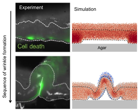 | 470 × 376 (189 KB) | Boaguilar (talk | contribs) |
- You cannot overwrite this file.
File usage
The following page links to this file: